15 Sep Overview of Musculoskeletal Ultrasound Scanning Service
Fast. Accurate. Non-Invasive.
Musculoskeletal ultrasound (MSK US) is a non-invasive diagnostic imaging technique used to assess soft tissue structures around joints, including muscles, tendons, ligaments, nerves, and joints themselves. Unlike traditional imaging modalities like MRI or X-ray, ultrasound uses high-frequency sound waves to produce real-time images, making it particularly useful for dynamic assessments.
This service is commonly used in sports medicine, orthopaedics, rheumatology, and physiotherapy, aiding in both diagnosis and treatment planning for a variety of musculoskeletal conditions.
Benefits of Musculoskeletal Ultrasound Scanning
1. Real-Time, Dynamic Imaging
Allows visualisation of movement, helping assess joint function, tendon gliding, and impingement in real time.
Enables guided injections and aspirations with high precision.
2. Non-Invasive and Safe
Uses no ionizing radiation, making it safer for repeated use and suitable for all patient populations, including children and pregnant individuals.
3. Quick and Accessible
Performed in clinic, allowing for immediate diagnosis and treatment decisions.
Faster than MRI.
4. Cost-Effective
Typically, more affordable than MRI or CT, offering a cost-efficient solution for both healthcare providers and patients. £125 at Ocean Physio & Rehab.
5. High-Resolution Imaging for Soft Tissues
Excellent for evaluating superficial structures such as tendons, ligaments, and small joints (e.g., hands, feet).
Capable of detecting subtle changes like small tears, inflammation, or fluid collections.
6. Patient Engagement
The patient can view the scan live, which can improve understanding of their condition and increase compliance with treatment plans. Report on the day of the scan.
What Musculoskeletal Ultrasound Is Not Suitable For
While Musculoskeletal Ultrasound is a powerful and versatile imaging tool, it does have limitations. It is not suitable for evaluating:
Deep joint structures such as the hip joint in obese patients or pelvis
Bone pathology, including fractures or bone tumours
Intra-articular cartilage damage, such as detailed assessment of menisci or labarum
Spinal conditions, including disc herniation or vertebral issues
Large or deep-seated soft tissue tumours (MRI is usually preferred)
Comprehensive full-body assessment
In these cases, other imaging methods like MRI, CT, or X-ray may be more appropriate. We’re happy to guide you or work with your healthcare provider to ensure you get the most accurate and appropriate diagnostics for your condition.

What We Scan
Shoulder (e.g., rotator cuff tears, impingement)
Elbow (e.g., tennis/golfer’s elbow)
Wrist & Hand (e.g., carpal tunnel, tendon injuries)
Hip & Groin (e.g., bursitis, tendinopathy)
Knee (e.g., ligament injuries, effusion)
Ankle & Foot (e.g., plantar fasciitis, sprains)
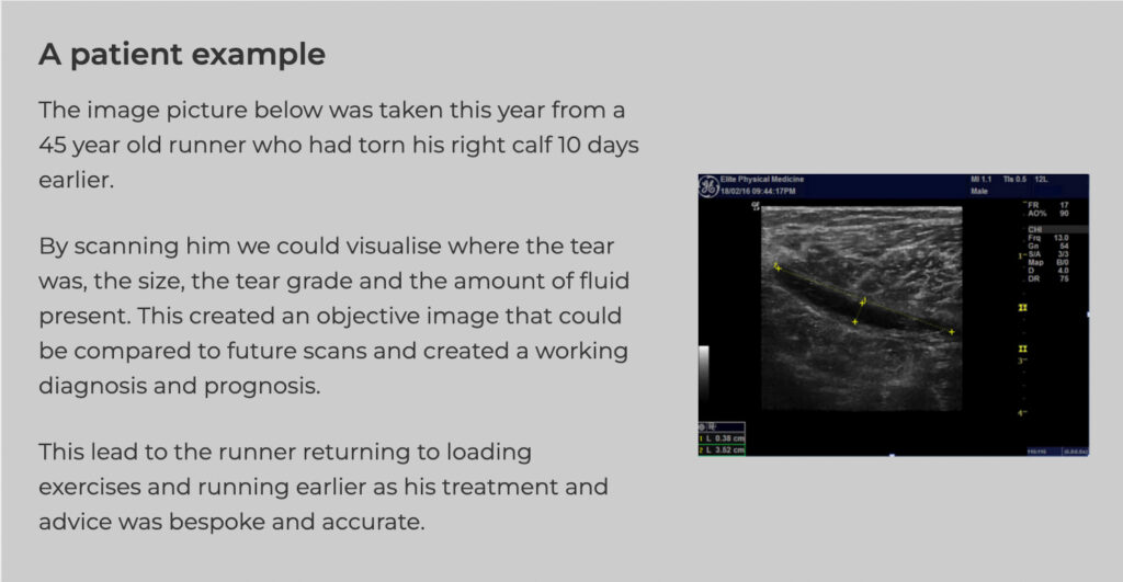
Muscle tears
Tendon pathology
Nerve entrapments and inflammation
Ideal For:
Athletes and active individuals
Patients with chronic joint or muscle pain
Post-operative monitoring
Suspected soft tissue injuries
GP or physio-referred diagnostics
